
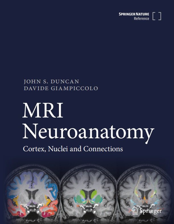
MRI Neuroanatomy: Cortex, Nuclei and Connections
01 Jan 2025, Springer Nature, ISBN Electronic: 9783031766961, ISBN Print: 9783031766954, English, 2883 pages
Writers: John S. Duncan & Davide Giampiccolo
About the book
The dogma that brain function relied on the cortex has dominated clinical neurology, neurosurgery and psychiatry for the last 100 years. Since the start of the 2000s, it has become evident that brain function is orchestrated as a network through white matter connections. This framework provides an understanding of brain function and dysfunction, and has radically changed how neurosurgical resections are performed. There is currently no manual for clinicians to visualise this functional anatomy in a fast, easy and user-friendly way. This is particularly important for senior clinicians who may have an understanding of cortical anatomy but may struggle with newly described white matter connections as they may now be visualised with MRI, and also for trainees who are learning the subject of applied neuro-anatomy. With this book, we aim to bridge this gap.
In 1995 Jackson and Duncan produced “MRI Neuroanatomy: a new angle on the brain” using the best clinical MRI available at the time and did not demonstrate white matter tracts. This was very well received, and it is time now to produce an atlas that shows the 3D anatomy of gray and white matter to contemporary standards.
This atlas is based on a high-quality MRI of a healthy subject which resembles the type of imaging is regularly available to clinicians. It is structured in sections (cortical anatomy, subcortical anatomy, and network anatomy) that are intended to guide clinicians from the classical cortical paradigms into a network neuroscience perspective. The 2D orthogonal slices are organised in two orientations:
(A) following the plane of the anterior and posterior commissure, as has been traditionally used in stereotactic atlases
(B) following the plane of the hippocampus, as is commonly used in clinical epilepsy practice.
The first part shows the 3D anatomy of the cerebral hemisphere, deep nuclei, brainstem and cerebellum.
The second part shows the same brain cut in 2D orthogonal slices (axial, coronal, sagittal) with a raw T1-weighted image, accompanied by a labelled image showing the gross anatomy of the brain and grey matter structures and, also, the labelled white matter tracts in that slice. This is particularly relevant for neurosurgeons, who will be able to appreciate before planning a resection the relationship between each tract’s trajectory and the gray matter. This will also benefit neurologists, enabling clarity as to how single lesions can cause multiple disconnection and impact on different functions and behaviours.
The third part demonstrates the 3D anatomy of the major white matter tracts in the brain, to indicate how distant lesions can impact the same function.
The book also contains
Detailed images of an individual brain showing detailed grey and white matter anatomy
Demonstration of inter-relationships between grey matter and white matter tracts and cortical and nuclear terminations
Atlas of brainstem nuclei and the relevant brainstem connections
Epilepsy Sparks Insights podcast episodes - with John Duncan
When Epilepsy Meds Fail - Where Neuromodulation Fits - Prof. John Duncan
-
John Duncan is a Consultant Neurologist specialising in epilepsy, practicing at the National Hospital for Neurology and Neurosurgery, Queen Square, London and at Chalfont Centre for Epilepsy, His personal research focus is neuroimaging applied to epilepsy surgery.
He was appointed Professor of Neurology at the UCL Institute of Neurology in 1998. From 2012 to 2018 he was Clinical Director of the National Hospital for Neurology and Neurosurgery. He is NIHR Senior Investigator and Governor of UCL Hospitals. He is Honorary Professor of Neurology, Fudan University, Shanghai, China. Visiting Professor, Faculty of Life Sciences & Medicine, Kings College London.
In 2004, John received the annual Clinical Research recognition award of the American Epilepsy Society. He is past-President and past treasurer of the UK chapter of ILAE. In 2005 he was elected Ambassador for Epilepsy and to be a Fellow of the Academy of Medical Sciences. In 2025 he was elected to the European Academy.
-
Davide Giampiccolo MD, PhD is a Senior Neurosurgical Fellow in Functional Neurosurgery at the National Hospital for Neurology and Neurosurgery in Queen Square, a Principal Investigator in the Department of Epilepsy at the UCL Institute of Neurology and an Emerging Leaders fellow for the Epilepsy Research Institute UK. He qualified in Medicine and Surgery between the Charité – Universitätsmedizin Berlin, Germany and the University of Verona, Italy in 2015. He completed his training in Neurosurgery in Verona, Italy under Prof. Francesco Sala and Montpellier, France under Prof. Hugues Duffau in 2021. He previously studied white matter anatomy under Prof. Marco Catani at King’s College London in 2016. He has completed his PhD/fellowship in complex surgical epilepsy at the UCL Queen Square Institute of Neurology in 2025 under Prof. John Duncan.
His research focused on brain circuits combining white matter anatomy and direct white matter stimulation. Together with Prof. Duffau, he has revised language models by rediscussing functional anatomy for the arcuate fasciculus (Giampiccolo and Duffau 2022 Brain) and the inferior fronto-occipital fasciculus (Giampiccolo, Herbet and Duffau 2025 Brain). He researches also specific white matter tracts involvement in seizure networks, and showed that discrete white matter pathways disconnection (i.e. anterior thalamic radiation) directly impacts long-term seizure freedom in frontal lobe epilepsy (Giampiccolo et al 2023 Brain). Within the field of intraoperative neurophysiology, he has pioneered cerebello-cortical stimulation and white matter stimulation with SEEG. He has also first adapted cortico-cortical evoked potentials to intraoperative resection for language preservation during asleep neurosurgery.
Davide has received a number of awards from scientific and clinical communities including the Organisation for Human Brain Mapping (2016), the International Society for Intraoperative Neurophysiology (2018), the European Association of Neurosurgical Societies (2020), the British branch of the International League against Epilepsy (2025) and the American Epilepsy Society (twice, in 2024 and 2025). In 2019 he was the first non-North American to receive the Academy Award from the American Academy of Neurological Surgery. In 2024 he was awarded the prestigious Emerging Leaders fellowship by the Epilepsy Research Institute UK. He is the scientific co-Chair of the International Society for Intraoperative Neurophysiology. Davide is an investigator of the GB Prime trial (Chief PI H. Akram), the largest trial in the world using the Neuralink BCI for patients with paralysis. He is the editor together with Prof. John Duncan of MRI Neuroanatomy: Cortex, Nuclei and Connections, a white matter atlas published by Springer Nature as a Major Reference work.