
Brain scans
All of the brain scans listed below are functional neuroimaging techniques for mapping brain activity. Which, is pretty important when trying to identify epilepsy or indeed many other brain issues!
Some of the scans can be noisy (MRI, fMRI), the EEG needs your scalp to be scratched a bit (sounds strange right?!) and some need you to go into a little tunnel for a period of time (all scans bar the EEG and MEG).
However, these scans are incredible and play a key part in helping diagnose and treat epilepsy.
Join us on social
Computed Tomography (CT or CAT)
Having a CT: you lie down on a motorised bed which moves into a tube-like scanner. Here, narrow beams of x-rays are aimed at your body at different angles so that it shows scans of "slices" of your body! It can be used on any part of your body. The machine looks a bit like that of an MRI or fMRI but isn't nearly as deep when you lie down.
The person doing the scan for you is called a Radiographer.
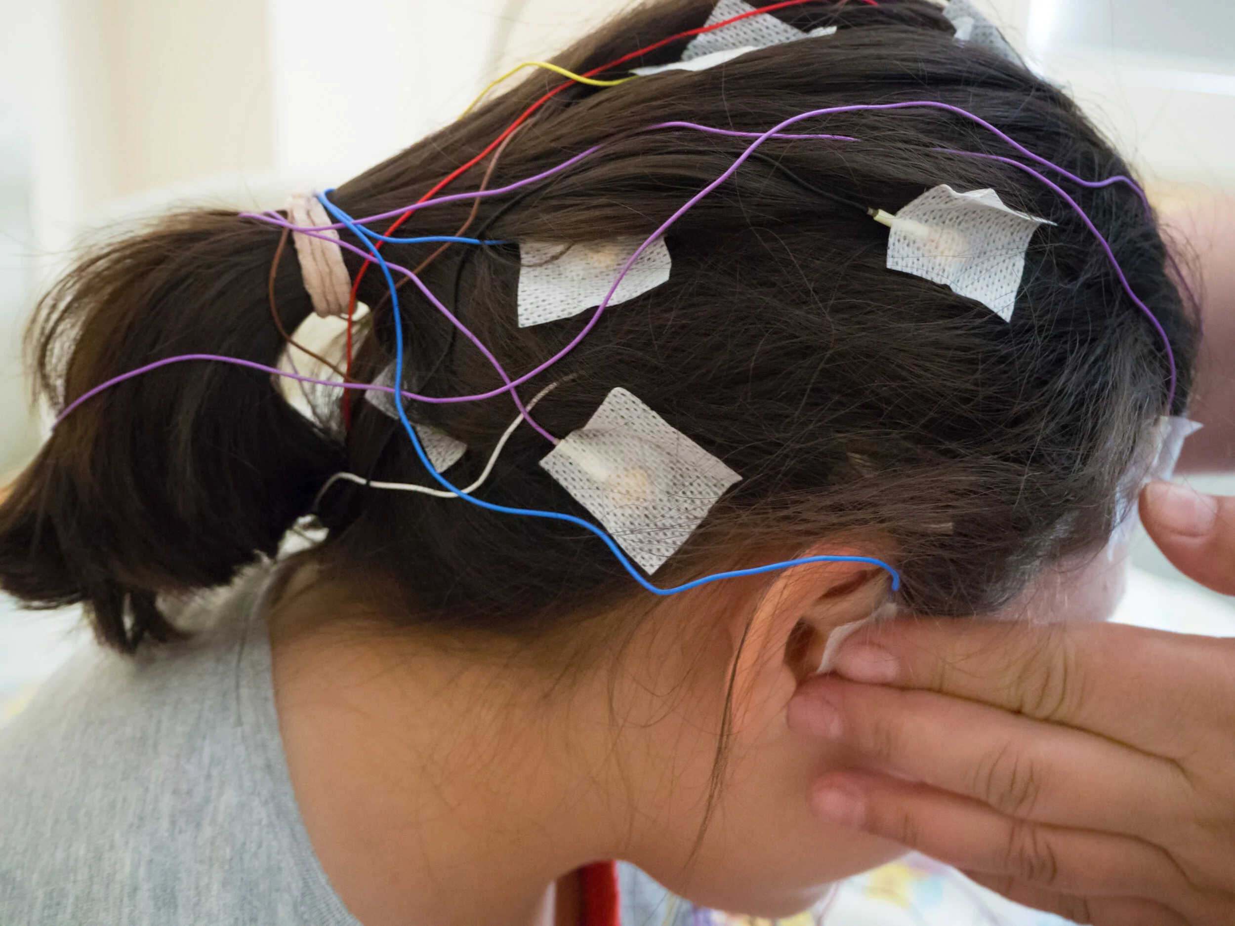
Electroencephalogram (EEG)
An EEG is where your brain waves are measured by electrodes. You sit down and have electrodes attached to your scalp in specific places. Your scalp will need to be scratched a little for good adherence of the electrodes. There are wires coming out of the electrodes which link to a computer to record your brain waves and hopefully any abnormal activity. It is often the first scan that a person suspected of having epilepsy has.
The person doing the scan for you is a Neurophysiologist.
Functional Magnetic Resonance Imaging (fMRI)
Having a fMRI: you lie down on a motorised bed which moves into a tube-like scanner which then scans your brain to detect information regarding the function and structure of your brain.
The person doing the scan for you is called a Radiographer.
Magnetic Resonance Imaging (MRI)
Having an MRI: you lie down on a motorised bed which moves into a tube-like scanner, which then scans your brain to detect information regarding the structure of your brain.
The person doing the scan for you is called a Radiographer.
Magnetoencephalography (MEG)
Is where you sit down in a comfy chair and place your head in a scanner (which is above you). It’s like a giant helmet but it doesn’t make any noise. It identifies brain activity and measures small magnetic fields produced in your brain to produce a Magnetic Source Image (MSI) and pinpoint the source of seizures. It can detect local brain activity with more accuracy than an EEG.
The person doing the scan for you is called a Radiographer.
Positron Emission Tomography (PET)
This is where you receive a radioactive injection (called a tracer) and then lie down on a motorised bed. The bed moves into a tube-like scanner.
It is used to figure out how your brain functions (or where it’s having problems functioning).
PET scans can be combined with CT scans to produce even more detailed images. These are known as a PET-CT scans.
The person doing the scan for you is a Radiographer.
Single Photon Emission Computed Tomography (SPECT)
This is where you receive a radioactive injection (called a tracer) and then lie down on a motorised bed. The bed moves into a tube-like scanner which will then move around you to produce detailed three-dimensional images.
It is mainly used to measure blood flow to and around your brain.
The person doing the scan for you is a Radiographer.