
How I Discovered Brain Art

Dana Simmons1 is a medical writer and science communicator in Chicago2, USA3. She holds a Ph.D. in neurobiology4 from The University of Chicago5. Her science-art1 has been exhibited around the United States and internationally, and she was a 2016 recipient of the New England Biolabs’ Passion in Science Award6.
Volunteering: Dana is Co-Editor-in-Chief7 of Science Unsealed8, the Illinois Science Council’s9 blog
Leisure time: Baking snickerdoodles10 and hiking volcanoes around the world

If you have epilepsy, you might see your brain as your enemy. It acts out when you least expect it. It steals your cognitive11 gifts and harms the body you’ve tried so hard to protect. For those who study the brain in laboratories, the squishy blob (yes - it squishes!) inside our heads can be a nuisance for other reasons. It’s complicated, confusing, and dumbfounding at times.
But if you take a step back from investigating it at the molecular level and really look at the brain one cell at a time, you’ll see that it’s actually quite beautiful.
How I discovered brain art
About six years ago, I was doing some experiments in the lab. I’m a neuroscientist12, and I was looking at brain cells under a microscope to see how they work. To see what was going on, I had to fill a single cell with glowing dye, and I had to wait about 40 minutes for the dye to fill up the whole cell. So, I went to get some tea. When I came back and looked under the lens, the cell had completely filled up, and all of its weaving branches were staring back at me through the lens of the microscope.
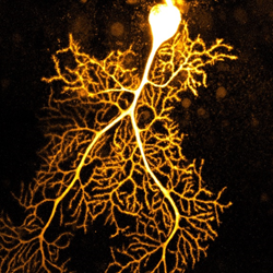

“Coral 2016” a Purkinje cell (14) - Dana Simmons (1)
This tiny cell (about two-tenths of a millimetre tall) looked like the most miniscule tree I’d ever seen. But it reminded me not only of tree branches, but also antlers, lightning, and coral. There was a pattern here. As I began to collect data, my mind kept coming back to the aesthetic appeal of these brain cells. Not only do they do amazing things that make us think, walk, balance, and make decisions, but they look rather snazzy too.

When the experiment ended, I took a picture of the cell and started adding colours, changing the lighting, and “art-ifying” the cell. It felt like I was taking portraits of these cells, adjusting the photos to showcase each of their uniquely branched shapes.
I began to think more about the pattern of branches and how it appears in so many parts of nature - microscopic and macroscopic. What was it about this pattern that made it so favourable throughout the test of time? It seemed to me that there must be an advantage to such a shape if it had formed in so many different parts of nature again and again. It was too much to be a coincidence. Was there a particular efficiency about it? Perhaps it had to do with management and delegating tasks? To have age-old corals share the structure with our phone-tree networks and decision diagrams....there just seemed to be more behind it.
Autism & the cerebellum
In any case, the scientific goal of my research was to learn about how autism16 changes the brain. I studied how cells communicate with each other in an autistic brain, and how that was different from a non-autistic brain. When most people think of autism, they think of changes in communication and social behaviour. However, a lot of people with autism also have changes to their movements - walking, balance, and eye movements. My research focused on the part of the brain that is involved with walking, balance, and eye movements.
As you might already know, many people with autism also have seizures17 - but not really in the part of the brain I studied. Seizures are more common in the cortical parts of the brain - behind your forehead, over your ears, the back of your head, and right on the top of your head. You see, these parts of the brain have a completely different structure from the part I studied. The part I studied, the cerebellum18 , has rigid, almost crystallized, architecture. It’s super organized, as if Marie Kondo19 popped into your brain and did some serious tidying. In contrast, the frontal and outer “cortical”20 parts of the brain...well, they do have some organization, but by comparison they’re rather jumbled - even in healthy, or neurotypical21, people.

The Cerebellum (22) – EnCor Biotechnology Inc (23)

The Cortex (24) – Frontiers in Neuroanatomy (25)
These cortical parts of the brain developed much later in evolution than the cerebellum, and they look enormously different. The individual cells are different shapes, and their networks are different shapes too. It seems that the structure of cells and networks in the frontal parts are quite a bit more prone to seizures because the waves of activity can propagate. The cerebellum just isn’t set up the same way. If you have epilepsy, I suppose you could think of the cerebellum as your champion brain region, since it’s unlikely to be affected by your seizures.
In case you’re curious, there have been documented cases of two people who had seizures connected with their cerebellums26. But still, this is only two people. It was so rare that their doctor actually wrote a research paper about these two patients. For context, at least 65 million people in the world live with epilepsy27.
What really fascinates me is to see how art and science combine so naturally. Regarding epilepsy, we can photograph brain cells using high-powered microscopes and turn the images into art. But it’s more than that. Art can give people a way to express how science affects them. Science is based on dry facts, which means that it is what it is - no more, no less. It remains objective, and necessarily so. Art, however, presents a venue for people living with epilepsy to show how their seizures feel and how they affect their life on a personal level. Art takes the human experience into account. Steven Schachter28, a professor of neurology at Harvard Med collects his patients’ art29. Many of his patients used art to explain how they felt when they were conscious during a seizure. Art is a fantastic, therapeutic outlet for lingering thoughts, worries, anxiety, and it provides a way to express yourself when words fall short.
Dana Simmons shares her passion for sharing the beauty of science and the complexity of the human neural system through photography. Learn more about the Passion in Science Awards™ at https://www.neb.com/about-neb/passion-in-science-awards
Trees Inside YourBrain: Exploring the Purkinje Pattern (30) - Dana Simmons (1)
Have you created art that depicts an element of your experience with epilepsy? What do you find most expressive? What inspires you?
